The medical industry has made large advancements since the beginning of society. Vaccines have made it possible to say goodbye to specific diseases. We’re all living longer and healthier lives thanks to modern medicine.
Radioisotopes are one aspect we need to thank. Since their discovery in the early 1900s, researchers have been finding more ways to use them. So, how are radioisotopes used in medicine today? That’s an excellent question, one we briefly delve into below. Keep reading to explore the world of radioisotopes and nuclear imaging.
What Is a Radioisotope?
Radioisotopes are species of chemical elements formed through the natural decomposition of atoms. Radioisotopes are also known as radioactive isotopes. The medical industry highly regards radioisotopes, even though radiation exposure is typically harmful; however, using radiation in specific applications is critical to diagnosing and treating diseases.
How Are Medical Radioisotopes Made?
Scientists can use two methods to create medical radioisotopes. One technique is flooding materials with neutrons in a reactor. They can also form from protons in an accelerator known as a cyclotron. A critical part of radiopharmaceuticals is radioisotopes. Some medical facilities have cyclotrons on campus to create radiopharmaceuticals with short-lived lives.
More About Radiopharmaceuticals
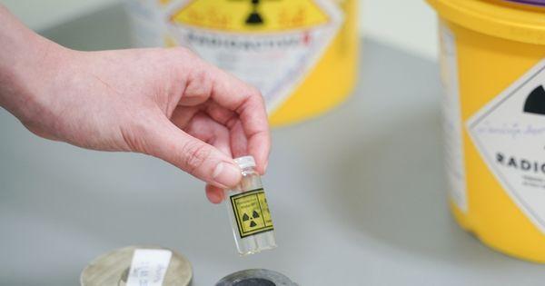
A radiopharmaceutical is a pharmaceutical molecule with a radioisotope tracer attached. Each patient is different; therefore, medical professionals carefully select and administer the proper amount of radiopharmaceuticals to ensure the patient’s safety. The pharmaceutical will gather in tumor tissue or a particular organ after entering the body. The attached radioisotope will start breaking down and producing specific radiation amounts used to treat injuries and diseases or diagnose an individual.
These tracers, either injected or taken orally, will allow various imaging methods to capture the emitted radiation. These techniques include positron emission tomography and single photon emission computed tomography. These images enable physicians to inspect blood flow to particular organs. They’ll also examine bone growth and organ function. Radioisotopes start decomposing before the radioactivity can cause harm to protect patients.
Common Radiopharmaceuticals
The most common radiopharmaceutical worldwide is technetium-99 (Tc-99). Upwards of 40 million procedures per year utilize this radioisotope for diagnosis.
Diagnostic Applications
The first diagnostic application we’re going to explore is nuclear imaging. This diagnostic technique uses radioisotopes that emit gamma rays from within the body. Nuclear imaging differs from other medical imaging systems, such as x-rays, MRIs, and CTs. The key difference is that the source of produced radiation is in the body via nuclear imaging. Medical professionals can see the radioisotope’s exact location, position, and concentration.
There are “hot spots” and “cold spots” within nuclear imaging. A “hot spot” indicates excess radioactivity around an organ or within the tissue that could be in a diseased state, like cancer or an infection. A “cold spot” will indicate blood isn’t getting through. Both of these issues will show up with nuclear imaging.
You may be wondering how nuclear imaging works. Patients receive radiopharmaceuticals as an injection, a drink, or something to inhale. A gamma camera then detects the drug and creates an enhanced image for the physician to view. Nuclear imaging discovers the body’s functions; it doesn’t provide anatomical photos of the body. It measures the accumulation or distribution of the radioisotope and blood flow.
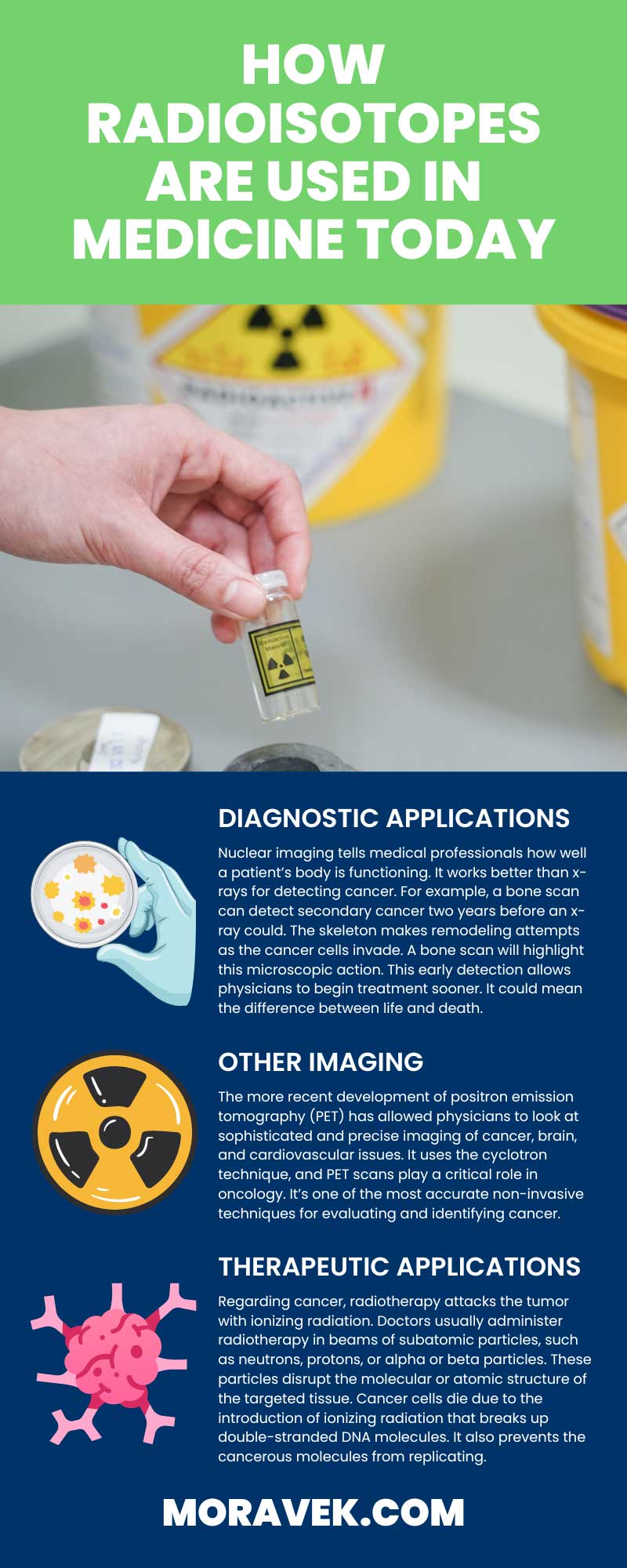
Nuclear imaging tells medical professionals how well a patient’s body is functioning. It works better than x-rays for detecting cancer. For example, a bone scan can detect secondary cancer two years before an x-ray could. The skeleton makes remodeling attempts as the cancer cells invade. A bone scan will highlight this microscopic action. This early detection allows physicians to begin treatment sooner. It could mean the difference between life and death.
Other Imaging
The more recent development of positron emission tomography (PET) has allowed physicians to look at sophisticated and precise imaging of cancer, brain, and cardiovascular issues. It uses the cyclotron technique, and PET scans play a critical role in oncology. It’s one of the most accurate non-invasive techniques for evaluating and identifying cancer.
Some medical professionals will combine PET scans with MRI or CT scans to provide even more precise results. Nuclear imaging is all about saving lives.
When you receive a PET and CT scan, you’re receiving a 30 percent more accurate diagnosis than one scan alone. It’s a vital tool for analyzing a variety of diseases, such as many types of cancer and dementia.
Combining PET and MRI scans is especially critical for brain imaging. It provides diffusion-weighted imaging in soft tissue with magnetic resonance spectroscopy and dynamic contrast.
Nuclear imaging is the key to saving lives. It positions the radiation source inside the body, rather than outside, for more precise results. Medical professionals can provide patients with correct diagnoses instead of months or years of misdiagnoses. It’s becoming more common for doctors and other physicians to ask patients to receive nuclear testing. They speed past the x-ray and other common scans to receive direct results.
Therapeutic Applications
Radioisotopes in therapeutic applications intend to destroy targeted cells. Medical professionals use radiotherapy to treat cancer and other abnormal growth tissue conditions, such as hypothyroidism.
Regarding cancer, radiotherapy attacks the tumor with ionizing radiation. Doctors usually administer radiotherapy in beams of subatomic particles, such as neutrons, protons, or alpha or beta particles. These particles disrupt the molecular or atomic structure of the targeted tissue. Cancer cells die due to the introduction of ionizing radiation that breaks up double-stranded DNA molecules. It also prevents the cancerous molecules from replicating. Yes, there are challenging side effects, but this method will effectively slow cancer progression. It can even prompt the regression of malignant diseases.
Progress in the Field
Since the discovery of radioisotopes in the early 1900s, the industry has been advancing. Neutrons that bombard stable elements are artificial radioisotopes. After this discovery, researchers dove head first into studying potential medical applications of artificial radioisotopes. This research laid the groundwork for nuclear medicine. In fact, medical professionals use therapeutic and diagnostic procedures routinely.
As you can see, professionals use radioisotopes quite often in medicine development today. The medical industry has made strides in the past century to better understand these hidden deadly diseases. One thing radioisotopes have clearly become synonymous with is cancer. It’s crucial to finding cancerous cells and curing them.
If you want a highly-qualified partner to create your radiolabeled pharmaceuticals, turn to Moravek, Inc. We’re GMP manufacturers with over 45 years of industry experience. We’ll meet your specific requirements and coordinate with you for a timely release of your product.